


Medical
About Us
![]()
Our Fields
Sustainability

Medical

Environment

Social


Our Fields
Serum amyloid A (SAA) is an apolipoprotein associated with HDL, and the molecular weight is 11.4 kDa. The protein has been evolutionarily conserved among a wide range of vertebrate species [*1]. And more anciently, SAA has been identified in an echinoderm, sea cucumber [*2].
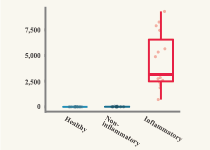
Data of horse SAA.
Created based upon data from: Jacobsen, S., Vinther, A.M., Kjelgaard-Hansen, M. et al. Validation of an equine serum amyloid A assay with an unusually broad working range. BMC Vet Res 15, 462 (2019).
SAA is also known as one of the acute phase proteins (APP) in serum and plasma.
Tissue damage, such as trauma, infection, stress, neoplasia and inflammation, triggers acute phase response (APR) as a part of the early-defense or innate immune system [*3]. APP are synthesized from liver and certain extrahepatic tissues during APR [*3, 4].
In some species, SAA is classified as a major APP (the concentrations increase more than 10-fold when inflammation occurs). For example, equine, feline, bovine, canine, goat, mouse, porcine, rabbit, sheep, cheetah, and elephant seal are found to have SAA as a major APP [*3, 5, 6] . The concentration of equine SAA increase from 10 to often 1000 times [*4].
Also, the change of SAA concentrations can provide important prognostic value. For equine SAA, the response time is 6-12 hours and the peak is 48 hours after inflammation starts [*4, 7]. For other species, generally SAA starts increase at 8-12 hours after inflammation occurs, and it reaches maximal levels in 24-48 hours [*8]. As inflammation is resolved, SAA concentration declines in a short term. Since the half-life period of SAA is estimated that 24-48 hours, the concentration returns to an undetectable level after synthesis ceases [*4, 9].